Microscopy
Microscopy Unit
Centro de Investigación del Cáncer (CSIC-USAL).
Campus Universitario Miguel de Unamuno s/n E-37007 Salamanca (Spain)
+34 923 293 16
The CIC-IBMCC Microscopy and Cytometry Unit has tried during these last two years to improve their services. Accordingly, the facility has acquired a new super-resolution equipment that will allow to get new images with an unprecedented sharpness. In addition to the new service.
In this Facility we provide complete services for:
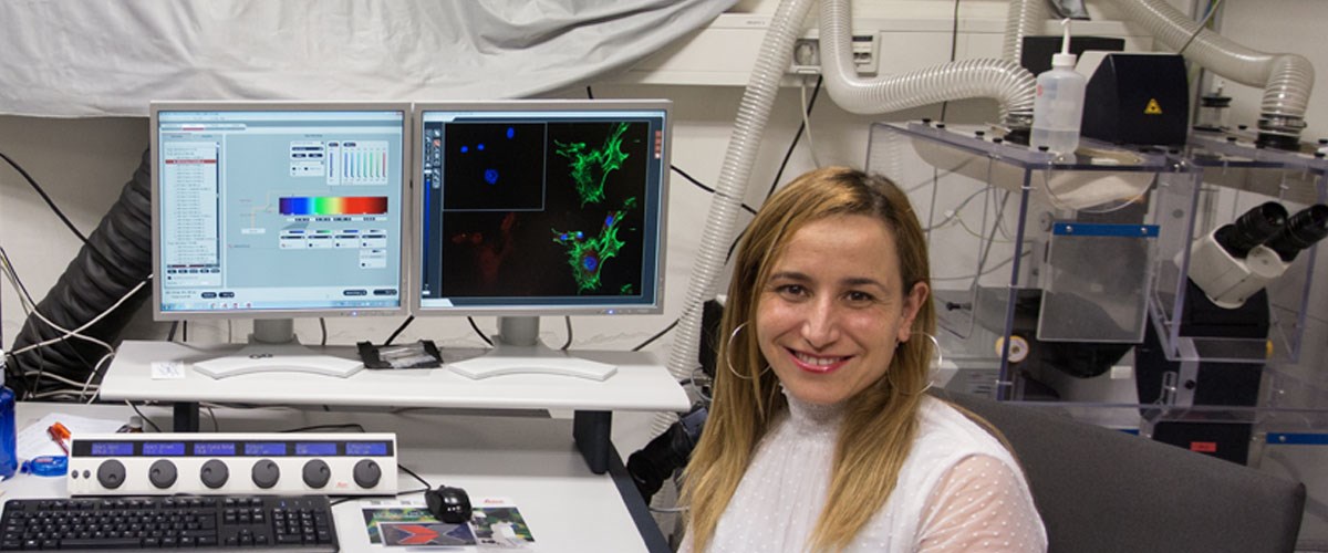
- Confocal microscopy (Leica SP5 and Zeiss LSM510), that provides services to internal and external users. Running timelapse, Z-Series, colocalization, FRAP and FRET experiments. Web based management allows remote communication with the service for reservations.
- Flow cytometry: sorting and immunophenotypic analysis of different cell populations, independently of complexity and/or staining. Running and maintenance of the BDFACS-Aria-III system. Configuration of FACSDiva software.
- Preventative and corrective maintenance of Accuri-C6 flow cytometer. Training and advising about Accuri-C6 use to the researchers so they can reach full autonomy. In addition, the Unit personnel prepare and refill the sheath solution and cleaning solutions when needed in order to keep the system in the optimum conditions.
- Live cell imaging: designing timelapse experiments and processing images and videos. Running Olympus IX71 and Nikon Eclipse TE2000 microscopes, both with CO2 and temperature controlled, and the software attached-Delta-Vision and Metamorph. Deconvolution of images available upon request.
- Conventional microscopy: 9 fluorescence microscopes, 10 inverted microscopes for cell culture, 1 microinjector, 1 microscope for cytogenetic and 1 microscope for histological analysis.
- Monthly revisions of the microscopes, including phase adjustment, Kohler adjustment, objective cleaning and weekly revisions of the different microscopy rooms (material reposition). Adjustment and maintenance of mercury and halogen lamps.
- Training and advising about image capture software and hardware (Metamorph, Leica LAS AF, LSM Image Browser, ImageJ, penlab, etc.). Solving inquiries about image analysis.
- Creating, updating and maintaining the Web Unit, including updates of technical specifications or new equipment incorporated by the Center. Edition of guides and tutorials about de instructions, basic configuration and software.
- Quality assessment. Certifications: ISO-9001 and Ohsas-18001.
Equipment
The Microscopy Unit of the Cancer Research Centre with its variety of equipments offers a high range of possibilities. There
are seven main equipments:
-
Laser Scan Confocal Microscopy Leica SP5: the machine was funded by FEDER, Ministerio de Sanidad y Consumo and Instituto de Salud Carlos III. The microscope has four lasers with seven excitation lines, which in combination with the spectral detection technology, allows any fluorochrome to be detected in the visible range. Due to its confocal module, it is highly demanded to obtain high resolution images of cell cultures or tissues. Aditionally, due to its software and the motorized stage, FRAP, FRET or co-localization analysis can be performed as well.
-
Flow cytometer and sorter BD FACS-AriaIII: co-funded by CSIC-MINECO and FEDER. It is one of the most advanced cytometry system available nowadays. It supports up to 6 excitation lasers combined with 20 detectors. This feature allows the staining with complex dye configurations. Furthermore, the cell damage is minimized by a temperature controlled system, increasing cell viability and process efficiency.
-
Live Cell Microscopy Delta-Vision: acquired with FEDER and CSIC founding, this equipment is mainly used for in vivo time-lapse experiments. The microscopy main advantages are the ultra-precise stage; the lighting system which combines a xenon lamp with an excitation filters wheel; and a high sensitive CCD camera. Thus, low exposition times are required reducing cell damage. In addition, the deconvolution module contributes to a more complex image processing.
-
Laser Scan Confocal Microscopy Zeiss LSM 510: mainly used when the demand is over possibilities of Leica confocal and for external users.
-
Microscope Nikon TE2000 for in vivo analysis: this microscope is combined with Metamorph, a very powerful software tool in microscopy. Metamorph provides several applications such as transforming images or quantization.
-
Cell analyzer cytometer BD Accuri-C6®: co-founded by CSIC-MINECO and FEDER. Despite Accuri-C6® is a simple and easy to use platform, it offers an optimum configuration which detects four fluorochromes simultaneously. Researchers can perform immunophenotipycal analysis, cell-cycle analysis, etc. Furthermore, different cellular models can be used, from yeast to mammals.
-
Laser Scan Confocal Microscopy Leica SP8: the equipment was co-founded by FEDER, Ministerio de Economía, Industria y Competitividad and Universidad de Salamanca. The microscope has a powerfull white laser that is able to excite ever wavelength into a width range. This, in combination with the spectral detection technology, allows any fluorochrome to be detected in the visible range. In addition two lasers can make the fluorescence depletion and the system provides hi-resolution images. Using this technology we can decrease the microscopy resolution limit to 50 nanometres. Moreover, the equipment is completed with a Huygens Deconvolution Software. The Huygens deconvolution provides a significant increase in the contrast and resolution, which significantly improves the visualization and analysis.
-
A Thunder® wide-field microscopy system with inverted microscope for in vivo experiments equipped with a TIRF (evanescence illumination), opto-genetic and photo-manipulation modules for FRAT, FRET, photo-conversion and uncaging experiments and high intensity lasers for single molecule localization experiments PALM/STORM and computational clearance, This equipment was acquired by the infrastructure support EQC2019-006002-P financed by MCIN/AEI/1013039/501100011033 and "FEDER A Way fo Making Europe".